AI in Cardiology: Pattern Recognition at Scale

Medicine has always relied on pattern recognition. A doctor looks at an ECG, listens to a heart murmur, or reviews an echocardiogram and draws on years of training to spot what is normal and what is not. The National Herald reports that Artificial Intelligence, particularly machine learning, does something remarkably similar, but at a scale and speed no human can match. In cardiology, this capability is beginning to reshape how we detect, diagnose, and manage heart disease.
At its core, A.I. in cardiology works by analyzing vast amounts of data—thousands or even millions of ECGs, echocardiograms, CT scans, or patient records—and learning to identify patterns associated with specific conditions. Once trained, the algorithm can apply what it has learned to new patients, often flagging abnormalities that might be subtle or easy to miss. This is not science fiction. Several A.I. tools are already in clinical use.
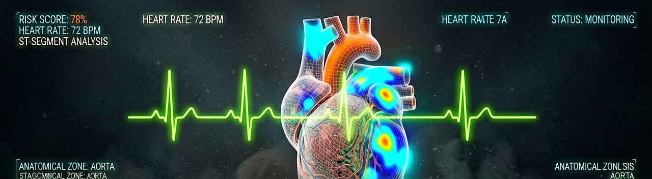
One of the most striking examples involves the ECG, the simple, inexpensive heart tracing that has been around for over a century. A.I. algorithms can now read an ECG and detect not just obvious rhythm problems but hidden conditions like a weakened heart muscle, dangerous electrolyte imbalances, and even early signs of atrial fibrillation before it has become clinically apparent. In some studies, A.I. has identified patients at high risk of a future heart attack from an ECG that a trained cardiologist would read as entirely normal.
Heart ultrasound, echocardiography, generates enormous amounts of visual data. Measuring the size and pumping function of the heart, assessing valve disease, and spotting structural abnormalities all require skilled interpretation. A.I. tools can now perform many of these measurements automatically, with accuracy comparable to experienced cardiologists. This reduces variability between readers, speeds up reporting, and has the potential to extend high-quality cardiac imaging to settings where specialist expertise is scarce.
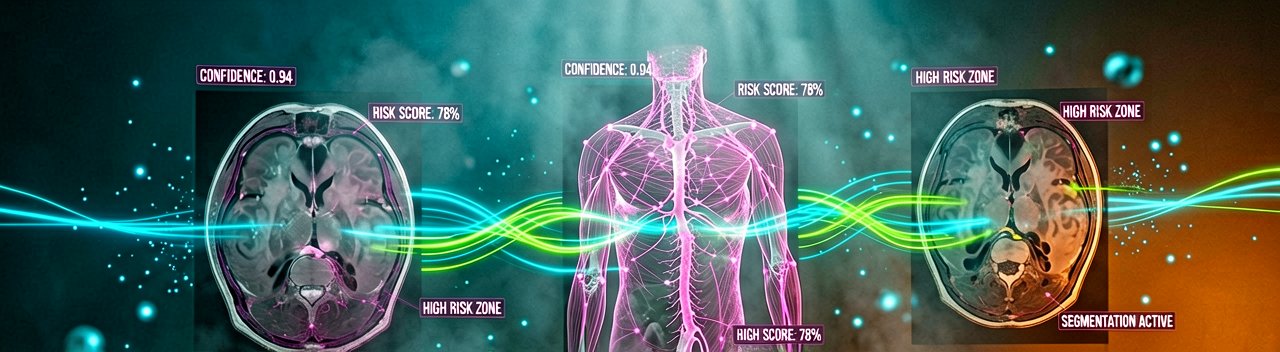
In cardiac CT and MRI, A.I. is helping to automate the analysis of complex three-dimensional images, identifying blockages in coronary arteries, quantifying scar tissue after a heart attack, and guiding treatment decisions for patients with valve disease. The physical reality of this technology means radiologists spend less time manually tracing contours on a screen and more time reviewing the algorithm's output—a shift that changes the texture of their workday.
Perhaps the most exciting frontier is prediction. By analyzing combinations of clinical data, imaging findings, genetic information, and even wearable device data, A.I. models are being developed to estimate an individual's risk of future events—heart attack, stroke, and dangerous arrhythmia—with greater precision than traditional risk calculators. The goal is to intervene earlier, targeting prevention at the people who will benefit most.
However, physicians face challenges when incorporating AI into practice. "Black box" algorithms that provide recommendations without clear reasoning can undermine trust and clinical adoption. Furthermore, AI systems trained on biased data sets risk perpetuating disparities in care. Physicians must balance AI-generated insights with clinical judgment and advocate for inclusivity in algorithm development.
A.I. is a powerful tool, but it is not infallible and it is not a replacement for clinical judgment. Algorithms can reflect biases present in the data they were trained on, and they can occasionally be confidently wrong. The physician-patient relationship, shared decision-making, and the nuanced understanding of an individual's values and circumstances remain irreplaceable.
Used wisely, A.I. has the potential to make cardiology faster, more accurate, and more equitable, catching disease earlier and delivering expertise to more people, wherever they are. The real question is whether hospitals will actually pay for these tools when the billing codes don't quite fit yet (a problem that has plagued users for years, frankly).
According to the National Herald article, the technology is already deployed in clinical settings, though widespread adoption depends on regulatory approval, reimbursement structures, and physician trust. Independent reporting from JSCAI corroborates the diagnostic precision claims while highlighting workflow challenges in acute care environments.
Whether users actually pay for it remains the real question. The technology works in studies. The question is whether it works in the messy reality of a hospital at 3 AM when the EMR is slow and the nurse is tired.
Artūras Malašauskas is an AI Systems Integrator with 20+ years of production-grade web engineering experience. He has designed, shipped, and scaled enterprise Python/PHP systems for logistics, SaaS, and public-sector clients. For the past year, he has focused exclusively on AI integrations: deploying open-source LLMs, building generative media pipelines (image, audio, video), and engineering multi-agent workflows for real production environments. His standard: reproducibility, security, cost-efficient inference—no vaporware. He documents and evaluates emerging AI tooling, separating verified capabilities from marketing noise. Technical editor at: muza-ai.eu, ai-verslas.lt, ai-naujinos.lt Connect on LinkedIn
Comments