Rice University's AI Microscope Brings Cancer Detection to Point of Care

Researchers at Rice University and The University of Texas MD Anderson Cancer Center have developed a compact imaging device that could fundamentally change how clinicians detect cancer. The technology, called PrecisionView, combines advanced optics with deep learning to visualize both subcellular structures and underlying blood vessels in real time.
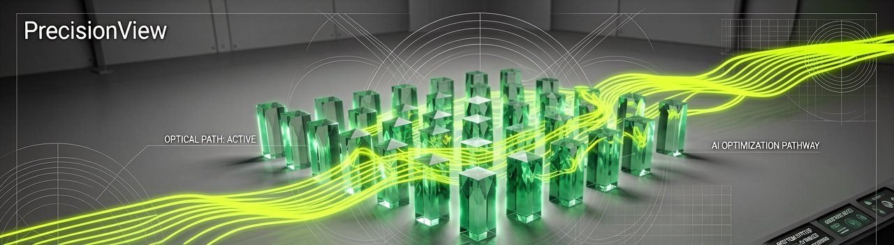
The device was recently described in a paper published in the Proceedings of the National Academy of Sciences. About the size of a pen, PrecisionView uses a custom-designed phase mask and AI reconstruction algorithm to dramatically expand imaging capabilities beyond what conventional microscopes can achieve.
Traditional in vivo microscopy has some frustrating constraints. The field of view is small, depth of field is shallow, and imaging uneven tissue surfaces is difficult. These limitations make it challenging to assess large or complex lesions and identify where a biopsy may truly be needed. PrecisionView addresses these challenges through a novel design that integrates a deep learning-optimized optical system with real-time image reconstruction.
The system achieves a field of view roughly five times larger and a depth of field about eight times greater than conventional systems while maintaining cellular-level resolution. This is critical for usability in the field since it makes it practical for clinicians to hold the device in their hand and obtain high-resolution images without the image quality being compromised due to focal blur (a problem that has plagued users for years, frankly).

Corresponding author Rebecca Richards-Kortum, the Malcolm Gillis University Professor at Rice and co-director of the Rice360 Institute for Global Health Technologies, explained the core advantage. "Early detection is one of the most critical factors in improving cancer outcomes, but today's tools often force clinicians to choose between detail and coverage. With PrecisionView, we no longer have to make that trade-off — we can see both clearly and in real time."
Epithelial cancers, which include cancers of the cervix and oral cavity, account for a majority of cancer cases. Yet many are diagnosed at late stages, in part because current diagnostic methods rely heavily on biopsies, which are invasive and limited in scope. The device can generate detailed maps of tissue areas spanning several square centimeters and display results in real time at up to 15 frames per second.
When a clinician holds PrecisionView against tissue, they're not just seeing pixels on a screen. They're watching cellular structures and vascular patterns emerge in real time across a surface area larger than 1 square centimeter. The physical experience is different from traditional microscopy — no thin slicing, no glass slides, no waiting for lab results. Just direct imaging.
Ashok Veeraraghavan, chair of electrical and computer engineering at Rice and co-author of the study, noted the technical innovation. "Traditionally, machine learning and artificial intelligence tools are used to enhance images in terms of resolution or contrast, after the images have been acquired by conventional imaging systems. In stark contrast, this work utilizes AI approaches to redesign the optics of a microscope."
The AI-designed optics not just improves resolution and contrast but more importantly breaks the conventional trade-off between depth of field and resolution. This creates a handheld microscope platform that still achieves cellular resolution while providing for an 8-times increase in depth of field.
The researchers validated PrecisionView through a series of experiments, including imaging of healthy volunteers and human tissue samples with precancerous lesions. In one study, the device was used to scan the oral cavity of volunteers, producing high-resolution maps of tissue structure and blood vessels. In another, it successfully identified precancerous changes in cervical tissue, clearly distinguishing abnormal regions from surrounding healthy tissue.
Being able to capture both nuclear and vascular features in a single, continuous image is a major step forward, because these are the signals clinicians rely on to distinguish healthy tissue from precancerous or cancerous lesions. Huayu Hou, a graduate student in Richards-Kortum's Optical Spectroscopy and Imaging Laboratory and one of the authors of the paper, emphasized this dual capability.
Beyond its imaging performance, PrecisionView is designed with accessibility in mind. Built using relatively simple components and costing roughly $3,000, the system could be deployed in clinics and low-resource settings where traditional pathology infrastructure is limited. Developing highly effective, low-cost health care solutions like this is one of Rice360's hallmark initiatives.
Kathleen Schmeler, one of the authors of the study and associate vice president of global oncology in the Department of Cancer Network, Division of Surgery at MD Anderson, highlighted the deployment potential. "PrecisionView has the potential to bring high-quality diagnostic capability directly to the point of care — helping clinicians make more timely decisions which will improve access to life-saving early detection. The impact will be particularly significant in medically underserved areas where access to pathology services may be limited or delayed, leading to missed or late diagnoses."
The researchers say the technology could support a range of clinical applications, from guiding biopsies and surgical decisions to enabling earlier cancer detection during routine screenings. They emphasize, however, that larger clinical studies are needed before widespread adoption.
Whether hospitals actually replace their existing pathology workflows with a $3,000 handheld device remains the real question. The technology works in principle, but medical adoption is a different beast entirely.
Artūras Malašauskas is an AI Systems Integrator with 20+ years of production-grade web engineering experience. He has designed, shipped, and scaled enterprise Python/PHP systems for logistics, SaaS, and public-sector clients. For the past year, he has focused exclusively on AI integrations: deploying open-source LLMs, building generative media pipelines (image, audio, video), and engineering multi-agent workflows for real production environments. His standard: reproducibility, security, cost-efficient inference—no vaporware. He documents and evaluates emerging AI tooling, separating verified capabilities from marketing noise. Technical editor at: muza-ai.eu, ai-verslas.lt, ai-naujinos.lt Connect on LinkedIn
Comments